
Fig. 2
- ID
- ZDB-FIG-141024-4
- Publication
- Xu et al., 1995 - Expression of truncated Sek-1 receptor tyrosine kinase disrupts the segmental restriction of gene expression in the Xenopus and zebrafish hindbrain
- Other Figures
- All Figure Page
- Back to All Figure Page
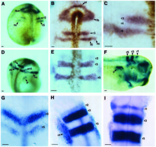
Expression patterns of XSek-1 and rtk1 in the developing hindbrain. (A-F) The expression pattern of XSek-1 during Xenopus development was analysed by whole mount in situ hybridisation. Photographs were taken of either cleared whole embryos (A,D,F), or of the neural epithelium after mounting under a coverslip (B,C,E). (A) Stage 14.5. (B) Rostral neural epithelium at stage 15. (C) Higher magnification view of hindbrain at stage 15. (D) Stage 20. (E) Hindbrain at stage 20. (F) Stage 33. (G-I) The expression pattern of rtk1 during zebrafish development was analysed by whole mount in situ hybridisation. Photographs from a dorsal view were taken after mounting of the hindbrain under a coverslip. (G) 11.5 h. (H) 17 h. (I) 24 h. r, rhombomere; fb, forebrain; nc, neural crest; ol, olfactory placode; ot, otic placode; p, pronephros. Scale bars, 50 µm. |
| Gene: | |
|---|---|
| Fish: | |
| Anatomical Terms: | |
| Stage Range: | 1-4 somites to Prim-5 |