
Fig. 5
- ID
- ZDB-FIG-141021-1
- Publication
- Hofmeister et al., 2013 - Distinct expression patterns of syndecans in the embryonic zebrafish brain
- Other Figures
- All Figure Page
- Back to All Figure Page
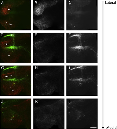
sdc4 expression surrounds the POC at 28 hpf. 2.0 µm optical saggital section taken through wholemounts of the zebrafish forebrain at 28 hpf from most lateral (A-C) to most medial (J-L). sdc4 expression (B, E, H, K and in red A, D, G, J) is shown in relation to the anterior and post-optic commissures and the neuronal clusters of the drc and vrc that contribute to these commissures respectively. Single sections taken laterally reveal the presence of sdc4 positive neuroepithelium lateral to the post-optic commissure (filled arrow in A). More medial sections reveal expression between the optic recess and POC (filled arrow in D-J) and also ventral to it in the presumptive hypothalamus (asterix, D-J). Abbreviations; dorsorostral cluster, optic recess (or), hypothalamus (hy), ventrorostral cluster (vrc). Scalebar in L is 25 µm. |
| Gene: | |
|---|---|
| Fish: | |
| Anatomical Terms: | |
| Stage: | Prim-5 |
Reprinted from Gene expression patterns : GEP, 13(3-4), Hofmeister, W., Devine, C.A., and Key, B., Distinct expression patterns of syndecans in the embryonic zebrafish brain, 126-32, Copyright (2013) with permission from Elsevier. Full text @ Gene Expr. Patterns