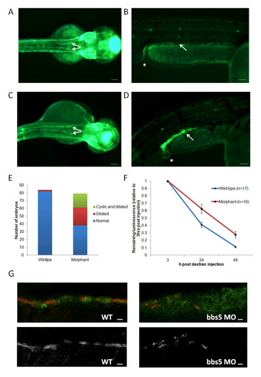
Characterisation of pronephros structure and function in bbs5 morphants. (A-D) Superior and lateral views of a cldnb:lynGFP embryo displaying (A,B) normal pronephric ducts (arrowed) and cloaca (*) in wildtype (WT) zebrafish. (C,D)bbs5 morphant embryos displayed dilated and tortuous pronephric ducts (identified by GFP fluorescence) and a (D) cloacal cystic dilatation. Scale bars are 100 μm. (E) Quantification of pronephros abnormalities in WT and bbs5 morphant embryos. (F) Estimation of GFR in zebrafish embryos was performed by measuring change in cardiac luminosity of both WT and morphant embryos after cardiac sinus fluorescent dextran injection. Mean luminescence +/- SEM (arbitrary units) is plotted versus time, up to 48 hours post injection. Morphant embryos retained significantly more fluorescent dextran at 24 hours (P = 0.005) and at 48 hours (P = 0.002). (G) At 72 hpf, compared to WT control, bbs5 morphant embryos revealed disrupted and fewer numbers of cilia in the dilated pronephros. Scale bar 10 μm).
|
