
Fig. 1
- ID
- ZDB-FIG-140822-7
- Publication
- Whitesell et al., 2014 - An alpha-smooth muscle actin (acta2/alphasma) zebrafish transgenic line marking vascular mural cells and visceral smooth muscle cells
- Other Figures
- All Figure Page
- Back to All Figure Page
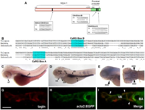
Acta2 promoter/enhancer construct design and expression in zebrafish. (A) A zebrafish (Dr) enhancer/promoter construct was constructed from the proximal promoter and first intron sequence of the zebrafish acta2 gene, and contains three highly conserved CArG binding sites also found in the mouse (Mm) acta2 proximal promoter and first intron. (B) Comparison of zebrafish CaRG boxes A and B in zebrafish, tilapia and medaka. (C,D) By wholemount in situ hybridization, acta2 shows strong expression in the gut (g) at 72 hpf (B), and expressed in the gut, swim bladder (sb), ventral aorta (va), floor plate (fp), aortic arch arteries (aaa), and bulbus arteriosus (ba) at 100 hpf (C). (E,F) Co-localization of wholemount in situ hybridization acta2 and anti-GFP staining of the acta2:GFP transgene shows strong expression in the aortic arch arteries (aaa) at 100 hpf. (G,H,I) 4 dpf acta2:EGFP transgenic fish (H) stained with Tagln rabbit polyclonal antibody (G). Merge (I) shows co-localization between acta2:GFP and Tagln. Arrowheads in G–I depict vascular mural cells. Scale bar in G represents 20 μm. |