
FIGURE
Fig. 5
- ID
- ZDB-FIG-140822-11
- Publication
- Whitesell et al., 2014 - An alpha-smooth muscle actin (acta2/alphasma) zebrafish transgenic line marking vascular mural cells and visceral smooth muscle cells
- Other Figures
- All Figure Page
- Back to All Figure Page
Fig. 5
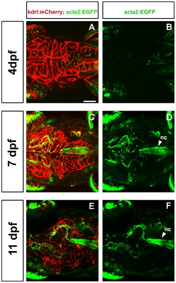
Development of mural cells and endothelial cells as seen in dorsal view. Confocal micrographs collected from a dorsal point of view show a progressive increase in vessel complexity (red, A, C, E) and in density of mural cell coverage of head vessels (green, B, D, F) at 4 dpf (A, B), 7 dpf (C, D), and 11 dpf (E, F). nc = notochord. Scale bar in A represents 100 μm. |
Expression Data
Expression Detail
Antibody Labeling
Phenotype Data
Phenotype Detail
Acknowledgments
This image is the copyrighted work of the attributed author or publisher, and
ZFIN has permission only to display this image to its users.
Additional permissions should be obtained from the applicable author or publisher of the image.
Full text @ PLoS One