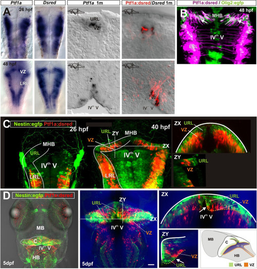
Distinct cerebellar progenitor populations are established early in the embryo. (A) Expression of ptf1a, DsRed (mRNA) and DsRed (protein) in developing and juvenile Ptf1a:DsRed transgenic fish. Ptf1a, DsRed and DsRed show similar expression patterns; (B) In vivo expression of DsRed and Egfp in the embryonic and juvenile Ptf1a:DsRed and Olig2:egfp transgenic fish. Overlapping Egfp and DsRed expression is seen in the VZ of the cerebellar primordium; (C) Nestin:egfp+ (green) and Ptf1a:DsRed+ (red) progenitors in the cerebellar primordium. The Nestin:egfp labels cells in the URL, while Ptf1a:DsRed line labels cells in the VZ. Two days post-fertilization Nestin:egfp+ and Ptf1a:DsRed+ cells form distinct populations in the URL and VZ of the cerebellar primordium; (D) A dorsal overview of Nestin:egfp+ (green) and Ptf1a:DsRed+ (red) progenitors in the hindbrain of a 5-day-old larval zebrafish. Two distinct progenitor domains are visible in the cerebellum (the junction is indicated with an arrow). Ptf1a:DsRed+ cells localize along the ventricular zone of the IVth ventricle (the VZ domain is labeled with a hatched orange line), while Nestin:egfp+ cells localize in the URL (hatched green line). MHB: Mid-hindbrain boundary; LRL: Lower rhombic lip.
|
