
Fig. S12
- ID
- ZDB-FIG-140527-46
- Publication
- Rydeen et al., 2014 - Cyp26 enzymes are required to balance the cardiac and vascular lineages within the anterior lateral plate mesoderm
- Other Figures
- All Figure Page
- Back to All Figure Page
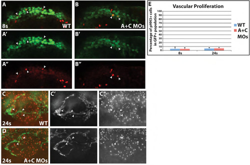
Vascular proliferation is not affected in Cyp26 deficient embryos. (A-B′′) Single optical section confocal images of WT and Cyp26 deficient TgBAC(etv2:EGFP) embryos at 8s immunostained for endothelial progenitors with GFP (green) and proliferation with pHH3 (red). Images are dorsal views of one side of the embryo with anterior to the right. (C-D′′) Single optical section apotome images of WT and Cyp26 deficient Tg(kdrl:nlsEGFP) embryos at 24 hpf immunostained for endothelial GFP (green) and pHH3 (red). Images are lateral view with anterior to the left. (E) Quantification of the percentage of proliferating vascular cells. At 8s, the WT vascular field had 4.4±4.3% proliferation compared to 4.3±2.9% in Cyp26 deficient embryos. At 24 hpf, the anterior vessels had 5.4±1.9% proliferating cells compared to 6.0±1.9% in Cyp26 deficient embryos. Arrowheads indicate proliferating VP cells. |