
Fig. S1
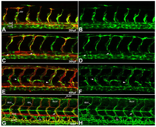
Expression pattern of flt4BAC:mCitrine reporter line at different stages of vascular development. (A-H) Double transgenic embryos for flt4:mCit (green) and kdrl:mCherry-Caax (red) at different stages of vascular development. (A, B) Initially, the flt4 reporter shows expression in both, arterial and venous ECs with an enriched signal within the venous compartment (26 hpf). (C, D) From about 26 hpf onwards, arterial expression of the construct decreases, so that emerging secondary sprouts can be easily followed at around 36 hpf (arrows). (E, F) At 2 dpf, the flt4 reporter expression is strongly confined to venous derived structures (venous ISV marked by arrowhead) and the signal gradually increases in the lymphatic lineage (see PLs highlighted by arrows). (G, H) By day 5, flt4:mCit expression is still evident in the PCV and venous ISVs (arrow). In addition, lymphatic structures including the TD, ISLVs as well as the DLAV are clearly highlighted throughout the trunk. [DA-dorsal aorta, PCV-posterior cardinal vein, TD-thoracic duct, ISV-intersegmantal vessel, ISLV-intersegmental lymphatic vessel, DLAV-dorsal longitudinal anastomotic vessel, DLLV-dorsal longitudinal lymphatic vessel] |