
FIGURE
Fig. 6
- ID
- ZDB-FIG-140512-24
- Publication
- Osborn et al., 2014 - Loss of FTO antagonises Wnt signaling and leads to developmental defects associated with ciliopathies
- Other Figures
- All Figure Page
- Back to All Figure Page
Fig. 6
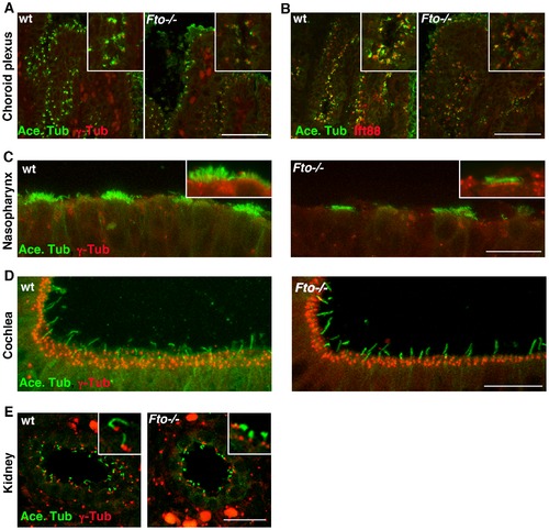
E15.5 Fto knockout mice embryos display tissue specific cilia defects. Paraffin sections from wild type and mutant animals showing immunolocalization of acetylated- α-tubulin (green) and γ-tubulin or IFT88 (red) in the choroid plexus (A,B); nasopharynx (C); cochlea (D); kidney (E). Loss of Fto results in shortened cilia in the choroid plexus, nasopharynx and kidney whilst cilia in the cochlea appear unperturbed. Scale bars: in A, B 50 μm; in C,D,E 20 μm. |
Expression Data
Expression Detail
Antibody Labeling
Phenotype Data
Phenotype Detail
Acknowledgments
This image is the copyrighted work of the attributed author or publisher, and
ZFIN has permission only to display this image to its users.
Additional permissions should be obtained from the applicable author or publisher of the image.
Full text @ PLoS One