
Fig. 5
- ID
- ZDB-FIG-140512-23
- Publication
- Osborn et al., 2014 - Loss of FTO antagonises Wnt signaling and leads to developmental defects associated with ciliopathies
- Other Figures
- All Figure Page
- Back to All Figure Page
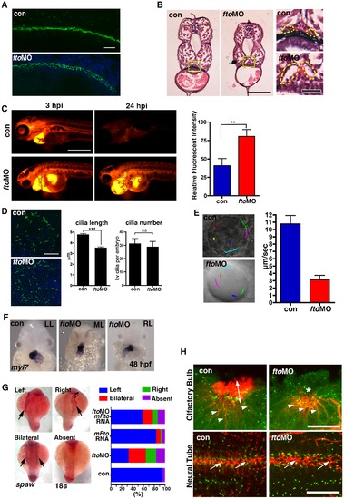
Loss of Fto in zebrafish leads to developmental defects associated with cilia dysfunction. (A) Cilia in the pronephric ducts of fto morphants are disorganised compared to wild type controls at 24 hpf. Cilia are marked by anti-acetylated tubulin (Green) and nuclei by DAPI (Blue); n = con 40/40, ftoMO 38/40 (B) Haemotoxylin and Eosin staining of wax sections through the pronephric ducts (at the level of the yolk extension) of control and fto morphants at 72hpf. Fto morphants develop dilated pronephric tubules, consistent with cilia disorganisation, highlighted by dotted lines in high magnification images to the right of the panel. Yellow squares demark magnified areas. Scale bars: 100 μm and 20 μm, low and high magnification images respectively. n = con 10/10, ftoMO 10/10. (C) Morphants have defective renal filtration as assayed through rhodamine dextran clearance assay. Images to the left of the panel depict embryos 3 hours post injection of the fluorescent tracer into the pericardium, the right side panels show the same fish 24 hpi, fto morphants retain significantly more fluorescence compared to controls, quantified and displayed as relative fluorescent intensity in graphical format (con: 40.98± SEM 9.539, n = 10. ftoMO: 80.54± SEM 9.516, n = 10. ** P<0.05). (D) Immunofluorecence of cilia, marked by anti-acetylated tubulin, and nuclei, DAPI, in the KV of 10 somite control and fto morphants. Morphants display shorter cilia than uninjected controls (con: 4.789±0.08773 N = 345, ftoMO: 3.557±0.06656 N = 464, *** P<0.0001) but have normal numbers of KV cilia per embryo (con: 31.36±3.757 N = 11, ftoMO: 29.00±3.896 N = 16, P = 0.6790, ns: not significant). (E) Fluorescent beads implanted into the KV at 12 hpf show that despite morphants displaying normal anticlockwise fluid flow, velocity of flow was significantly slower (con: 10.8 µm/sec ±1.1, fto MO: 3.2 μm/sec ±0.6, n = 5, movies in supplementary material). (F) In situ hybridisation for myl7 in control and fto morphants at 48 hpf show knockdown of Fto results in aberrant left-right patterning as observed by left looping (LL: 34/46), midline looping (ML: 5/46), and right looping (RL: 7/46) of the heart. (G) In situ hybridisation of spaw at 18 somite stage showing laterality associated defects in fto morphants that can be rescued when co-injected with mouse Fto RNA, see graph. (H) Acetylated- (red) and γ-Tubulin, marking the cilia and basal bodies respectively. FtoMO embryos show loss of cilia in the olfactory pit (upper panels). Arrow heads: olfactory neurons, double headed arrow: olfactory cilia, asterisk: reduced/absent olfactory cilia. Highly disorganised cilia in the neural tube (lower panels) of fto morphants at 24 hpf (arrows). Scale bar: 20 μm. N = con 10/10, ftoMO 23/31. |
| Fish: | |
|---|---|
| Knockdown Reagent: | |
| Observed In: | |
| Stage Range: | 5-9 somites to Protruding-mouth |