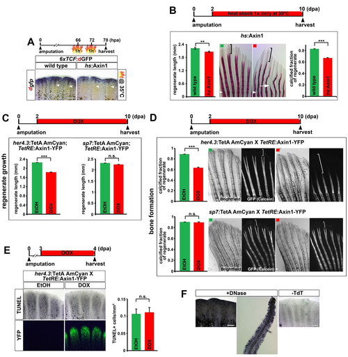
Fig. S4
- ID
- ZDB-FIG-140430-28
- Publication
- Wehner et al., 2014 - Wnt/β-Catenin Signaling Defines Organizing Centers that Orchestrate Growth and Differentiation of the Regenerating Zebrafish Caudal Fin
- Other Figures
- All Figure Page
- Back to All Figure Page
|
Inhibition of Wnt/β-catenin signaling in the proximal medial blastema interferes with regenerative growth and bone calcification, related to figure 4. (A) Mild heat shocks at 35°C strongly reduce proximal 6xTCF:dGFP Wnt reporter activity (arrowhead) but have little impact on distal Wnt reporter expression in 6xTCF:dGFP; hs:Axin1 double transgenic fish. (B) Systemic overexpression of axin1 at a level that has little impact on regenerative growth (daily heat shock at 35°C), strongly reduces the fraction of the regenerate containing calcified bones in hs:Axin1 transgenic fish. Bracket indicates the non-calcified distal region of the regenerate. Note that calcified bones are also thinner and shorter (arrow) compared to wild type control. (C) axin1 overexpression in the proximal medial blastema for 8 days reduces regenerate length in her4.3:TetA AmCyan; TetRE:Axin1-YFP double transgenic fish treated with DOX. axin1 overexpression in committed osteoblasts for 8 days has no effect on regenerate length in sp7:TetA AmCyan; TetRE:Axin1-YFP double transgenic fish treated with DOX. n = 9 fish, 6 rays each. (D) axin1 overexpression in the proximal medial blastema for 8 days in her4.3:TetA AmCyan; TetRE:Axin1-YFP double transgenic fish treated with DOX strongly reduces the fraction of the 11 regenerate containing calcified bones as determined by Calcein staining. axin1 overexpression in committed osteoblasts has no effect on bone calcification in sp7:TetA AmCyan; TetRE:Axin1-YFP double transgenic fish treated with DOX. Bracket indicates the non-calcified distal region of the regenerate. n = 9 fish, 6 rays each. (E) axin1 overexpression in the proximal medial blastema for 1 day does not enhance cellular apoptosis as detected by TUNEL assay in her4.3:TetA AmCyan; TetRE:Axin1-YFP double transgenic fish treated with DOX. (F) Positive and negative control for the TUNEL assay shown in (E). Longitudinal section of the DNAseI-treated positive control shows that TUNEL+ cells can be detected in the entire fin regenerate, including the osteoblasts. (B, D) Small arrowheads: amputation plane. (A, E-F) Scale bars: 200 μm. (B-E) Error bars indicate error of the mean. |

