Fig. S1
- ID
- ZDB-FIG-140430-25
- Publication
- Wehner et al., 2014 - Wnt/β-Catenin Signaling Defines Organizing Centers that Orchestrate Growth and Differentiation of the Regenerating Zebrafish Caudal Fin
- Other Figures
- All Figure Page
- Back to All Figure Page
|
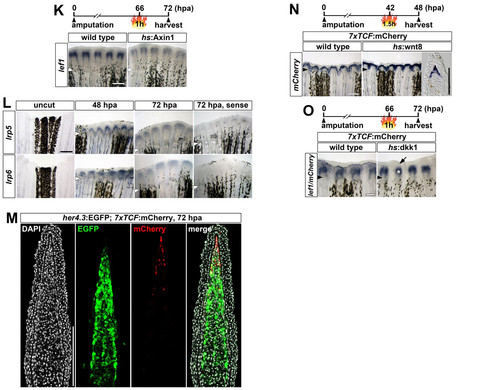
Wnt/β-catenin pathway activation, Tcf/Lef transcription factor and Lrp5/6 Wnt coreceptor expression during caudal fin regeneration, related to figure 1. (A) mCherry RNA expression in whole mounts of 7xTCF:mCherry transgenic regenerates. Note that transcript expression is detected at 6 hpa in the interray tissue (white arrowhead). (B) 7xTCF:mCherry reporter activity in the interray tissue (white arrowhead) is abolished upon dkk1 overexpression in 7xTCF:mCherry; hs:dkk1 double transgenic regenerates 6 hours post heat shock. (C) Confocal image showing mCherry fluorescence and Lef1 protein on sections of 7xTCF:mCherry transgenic regenerates. Note that mCherry fluorescence is not detected in the Lef1-positive basal epidermal layer. (D) mCherry fluorescence in 7xTCF:mCherry transgenic regenerates at different stages after amputation. Note that mCherry fluorescence is first detected in the interray tissue at 16 hpa. (E) Downregulation of axin2 expression in hs:Axin1 transgenic fish 6 hours post heat shock. (F) gfp RNA expression in Top:dGFP transgenic regenerates. Note that transcript expression is detected in the blastemal mesenchyme while the basal epidermal layer is devoid of signal (asterisk). (G) lef1, tcf1 (tcf7), tcf3a (tcf7l1a), tcf3b (tcf7l1b) and tcf4 (tcf7l2) are expressed in the regenerating fin. Hybridization with sense control probes revealed no signal. (H) Sections of whole mount stained regenerates shown in (G) reveal lef1 expression in the distal blastema (arrowhead) and the basal epidermal layer, tcf1 expression in the basal epidermal layer, lateral (asterisk) and distal blastema (arrowhead), tcf3a, tcf3b and tcf4 expression in the medial and lateral (asterisk) blastema. (I) tcf1 (brown) and lef1 (blue) are co-expressed in the epidermis of the regenerating fin. 5 (J) Dual color ISH of Tcf/Lef transcription factors and mCherry in 7xTCF:mCherry transgenic regenerates. tcf1, lef1 and tcf3a (blue) are co-expressed with mCherry (brown) in the distal blastema (black arrowheads), while tcf3b and tcf4 (blue) are not expressed in the distal mCherry (brown)-positive domain (white arrowheads). (K) lef1 expression is reduced in hs:Axin1 transgenic regenerates 6 hours post heat shock. (L) lrp5 and lrp6 are expressed in the regenerating fin. Hybridization with sense control probes revealed no signal. (M) Confocal image showing mCherry-positive and EGFP-positive cells on sections of 7xTCF:mCherry; her4.3:EGFP double transgenic regenerates at 72 hpa. (N) wnt8 overexpression does not cause ectopic Wnt reporter activation in the wound epidermis in 7xTCF:mCherry; hs:wnt8 double transgenic fish. (O) dkk1 overexpression interferes with mCherry (arrow) but not epidermal lef1 (asterisk) expression 3 hours after a single heat shock in 7xTCF:mCherry; hs:dkk1 double transgenic regenerates. (A-O) Small arrowheads: amputation plane. Scale bars: whole mounts, 200 μm and 100 μm (J); sections, 100 μm. |


