
Fig. 1
- ID
- ZDB-FIG-140422-15
- Publication
- Roberts et al., 2014 - Targeted transgene integration overcomes variability of position effects in zebrafish
- Other Figures
- All Figure Page
- Back to All Figure Page
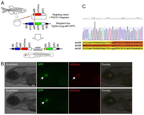
In vivo detection system for targeted integration of reporter constructs in zebrafish. (A) Schematic of PhiC31 targeted integration system. Transgenic embryos containing an attP docking site have a green lens due to the gamma-crystalline promoter driving a GFP reporter. ITR labels recognition sequences of either Tol2 or Sleeping beauty transposases used in generating the recipient transgenic lines with docking site. Injection of a circular plasmid with attB and a red reporter (targeting vector) into recipient line eggs leads to eye colour change upon PhiC31-targeted integration in larvae. (B) Detection of eye colour change upon PhiC31-mediated integration in the transgenic recipient Tg(Xla.crygc:attP-GFP)uobL6 line. Top row shows a transgenic larva with PhiC31-mediated recombination of the attB-mCherry cassette into the attP-GFP docking site. Bottom row shows transgenic sibling from the recipient line without targeted integration. Side views on cranial end of 5 dpf larvae with anterior to the left. Scale bar: 100 µm. Arrows indicate reporter expression in the lens. (C) Sequence of the tpl102 recipient docking site with germline integration of targeting construct tnnT2:attB-mRFP. Sequence shows the attR site in the crygc:attR-Red recombination product. The three lower case nucleotides denote the recombination site. |