
Fig. 2
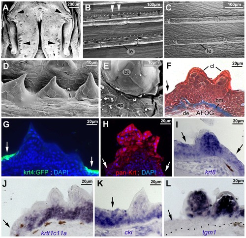
Breeding tubercle keratinocytes undergo more advanced keratinization. All images show breeding tubercles in wild-type zebrafish of one year of age; (A–E) views on body surface; (F–L) transverse sections through lower jaw. (A) Scanning electron micrograph (SEM) of the lower jaw; breeding tubercles are indicated by arrowheads. (B,C) Nomarski micrographs of pectoral fins of male (B) and female (C) fish. Bony rays (lepidotrichia) are marked (le). (D,E) SEM images of breeding tubercles (bt) on a male pectoral fin. (F) Trichrome (AFOG) staining; breeding tubercles consist of more cell layers than adjacent epidermis (arrow). The tubercle cap layer (cl) is stained in lighter red, resulting from its stronger keratinization. (G–L) GFP fluorescence of tg(krt4:GFP) line (G, green) and pan-type II Keratin immunofluorescence (H; red), in combination with DAPI staining. (I–L) in situ hybridization with indicated probes. Epidermis is indicated by arrows; dotted line in L marks basement membrane. Abbreviations: bm, basement membrane; bt, breeding tubercle; cl, cap layer; de, dermis; ep, regular epidermis; le, lepidotrichia. |
| Genes: | |
|---|---|
| Antibody: | |
| Fish: | |
| Anatomical Terms: | |
| Stage: | Adult |