
Fig. 3
- ID
- ZDB-FIG-140416-23
- Publication
- Staudt et al., 2014 - High-resolution imaging of cardiomyocyte behavior reveals two distinct steps in ventricular trabeculation
- Other Figures
- All Figure Page
- Back to All Figure Page
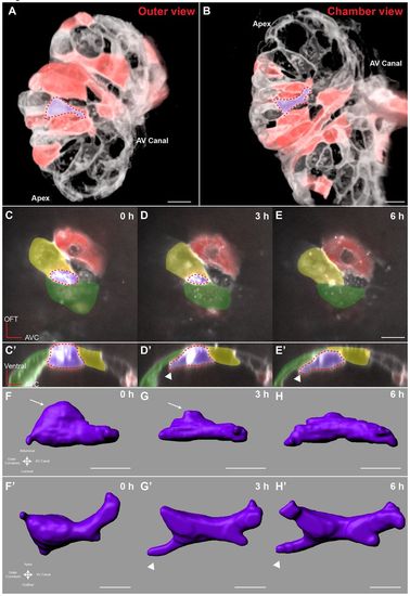
Cardiomyocytes show evidence of abluminal constriction. (A) 3D exterior view of a 72 hpf Tg(myl7:ras-GFP) heart with a subset of cardiomyocytes randomly labeled with RFP. Membrane GFP signal is pseudocolored white, and mosaically marked cardiomyocytes are in red. A cardiomyocyte of interest is highlighted in purple. (B) Interior chamber view of the same heart. Cardiomyocyte of interest is highlighted in purple. (C-E) Time-lapse highlighting a cardiomyocyte of interest in another heart. A single xy plane is shown at the start of imaging (C), at +3 hours of imaging (D), and at +6 hours of imaging (E). Orientation is as in A. Dotted lines outline the gradually constricting cell contact with the abluminal surface. The neighboring cardiomyocytes are highlighted in yellow and green. In E, the cardiomyocyte has lost contact with the abluminal surface and thus can no longer be seen. (C2-E2) Re-slice of the confocal stack showing the same cardiomyocyte rotated 90°. Cardiomyocyte of interest is highlighted in purple, and neighboring cardiomyocytes are in yellow and green. The cardiomyocyte can be seen extending a protrusion (arrowheads) as it exits the compact layer. (F-H) 3D volume rendering of the highlighted cardiomyocyte. Arrows point to contracting contact with the abluminal surface. Arrowheads point to the extending protrusion shown in E2. (F2-H2) 90° rotation of F-H showing luminal view of the cardiomyocyte. Scale bars: 10 μm in A-E; 5 μm in F-H2. |
| Gene: | |
|---|---|
| Fish: | |
| Anatomical Term: | |
| Stage: | Protruding-mouth |