acsl4a MOs specifically inhibit maternal acsl4a transcripts essential for dorsoventral patterning defect, related to Figure 1.
(A-D) Two translation-blocking acsl4a MOs dorsalize zebrafish embryos.
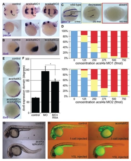
(A) Whole-mount mRNA in situ hybridization of pax2a in 20 hpf embryos of control, acsl4a MO1-injected (750 fmol) or acsl4a MO2-injected (750 fmol) embryos. pax2a marks m/h: midbrain/hindbrain boundary, r: anterior retina. (top) Dorsal view, anterior to the left. (bottom) Lateral view, anterior to the left. White arrowhead indicates wound-up trunk.
(B) tbx6, a marker of dorsal presumptive paraxial mesoderm, is expanded ventrally in acsl4a MO1- and acsl4a MO2-injected embryos (750 fmol). Lateral view, dorsal to the right.
(C-D) acsl4a MOs dose dependently decreases ventral marker gata1a.
(C) Representative images of whole-mount mRNA in situ hybridization of gata1a expression in embryos injected with increasing doses of acsl4a MOs. gata1a expression was binned into one of three levels: wild-type (blue), decreased (yellow), or absent (red). 8-somite stage embryos. Dorsal view, anterior to the left.
(D) Percentages of embryos with wild-type (blue), decreased (yellow), or absent (red) levels of gata1a expression after injection of varying doses of acsl4a MOs (acsl4a MO1: n=31-50 embryos, from 3 separate experiments; acsl4a MO2: n=30-60 embryos, from 3 separate experiments).
(E-F) Injection of acsl4a mRNA attenuates the acsl4a morphant’s dorsalized phenotype.
(E) Representative images of whole-mount mRNA in situ hybridization of tbx6 expression in 80% epiboly stage embryos. Embryos were uninjected (control), injected with acsl4a MO1, or injected with acsl4a MO1 combined with acsl4a mRNA lacking the MO target sequence. Vegetal pole view, dorsal to the right.
(F) Quantification of the angle of tbx6 expression. Data are mean ± SE of a representative experiment with more than 20 embryos per condition. ANOVA with REGWQ post-hoc test was performed. * p<0.05
(G-H) acsl4a is required maternally for dorsoventral patterning.
(G) MOs that inhibit splicing of zygotic Acsl4a transcript do not yield dorsoventral patterning defect. Representative bright field images of control embryo and embryo injected with acsl4a splice-site blocking MO (30 hpf; spliceMO1 1000 fmol).
(H) Acsl4a is not required in the yolk syncytial layer for proper dorsoventral patterning. Representative combined bright-field and fluorescent images of embryos injected with a fluorescein-conjugated MO (control) to indicate injection site or fluorescein-conjugated MO plus acsl4a MO (translation blocking acsl4a MO1). 30 hpf, lateral view, anterior to the left. (Top) Embryos were injected at the 1-4 cell stage. (Bottom) Embryos were injected into the yolk syncytial layer (YSL) at 1K-cell stage. The MOs were restricted from the cells of the embryo proper. 99% of embryos injected at the 1-cell stage with acsl4a MO were dorsalized in comparison to 3% of those injected in the YSL (n=92-93 embryos from 4 experiments). Embryos injected with control MO into the YSL also had occasional dorsalization (4%, n=52 embryos from 3 experiments), whereas 0% of control embryos injected at the 1-cell stage were dorsalized.
|
