
Fig. S4
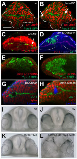
(A-B) wildtype and laminin-γ1 morphants, respectively, expressing H2b-RFP and Pard3- GFP. Arrows in (B) point at mis-positioned pard3-GFP puncta in the eye field of the laminin- γ1 morphant. (C) laminin-γ1 morphant immunostained as detailed in the panel. Arrows point at mispositioned ZO-1 puncta. (D) laminin-γ1 morphant cells (green) transplanted into a wild type eye field show no overt phenotype. Embryo immunostained as detailed in the panel. (E-F) frontal and dorsal views, respectively, of 3ss Tg(rx3:GFP) embryos showing laminin-β1 mRNA accumulation (red) and GFP protein (green). laminin-α1 and laminin-γ1 show similar expression patterns (not shown). (G-H) BSA-coated (G) and Laminin1-coated (H) beads implanted in the eye field. Embryos were left to develop until 10ss and immunostained as detailed in the panels. (I-L) Frontal views of 24hpf embryos of the genotypes detailed in the panels. |
Reprinted from Developmental Cell, 27(3), Ivanovitch, K., Cavodeassi, F., and Wilson, S.W., Precocious acquisition of neuroepithelial character in the eye field underlies the onset of eye morphogenesis, 293-305, Copyright (2013) with permission from Elsevier. Full text @ Dev. Cell