
Fig. S2
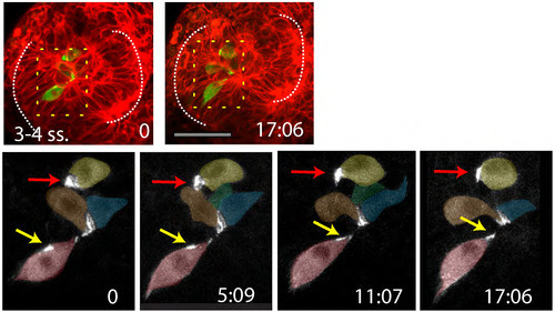
(A-B) 9ss (A) and 11ss (B) Tg(emx3:YFP) embryos immunostained as detailed in the panels. (Ai, ii, iii) and (Bi, ii, iii) show high magnifications of the telencephalic domain. ZO-1 is only weakly expressed at 9ss (Aii) but by 11ss, it is strongly expressed adjacent to the lumen of the telencephalon (Bii). (C) Snapshots from a time lapse movie of a pard3-GFP mRNA injected embryo, at 9ss (i), 11ss (ii) and 12ss (iii), showing the gradual appearance of polarisation in the telencephalon. (D) high-resolution detail of a telencephalic cell expressing lifeact-RFP and pard3-GFP, as it undergoes a midline cell division and its daughter cells establish apical domains at the telencephalic midline. White dotted lines in (A-B) outline the eye field; lines in (Ai-iii; Bi-iii; Ci-iii) outline the telencephalon. Vertical dotted lines in (D) highlight the future midline. |
Reprinted from Developmental Cell, 27(3), Ivanovitch, K., Cavodeassi, F., and Wilson, S.W., Precocious acquisition of neuroepithelial character in the eye field underlies the onset of eye morphogenesis, 293-305, Copyright (2013) with permission from Elsevier. Full text @ Dev. Cell