
FIGURE
Fig. 5
Fig. 5
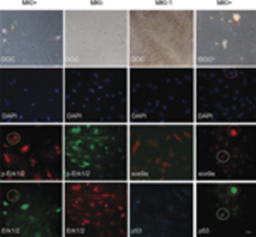
Involvement of MAPK-sox9a and p53 pathways in morphological alterations of cultured gonadal tissue. Ovary tissues were sliced and cultured in vitro. After 2–3 days, MKI or DMSO control were added and tissues were incubated for 48h. The cultures were costained with Erk1/2 and p-Erk1/2 antibodies (left two columns) or costained with sox9a and p53 antibodies (right two columns). DAPI staining and DOC were seen. Apoptotic or degenerating cells (arrow) and related signal staining (dotted circle) are shown. Scale bars=20μm |
Expression Data
Expression Detail
Antibody Labeling
Phenotype Data
Phenotype Detail
Acknowledgments
This image is the copyrighted work of the attributed author or publisher, and
ZFIN has permission only to display this image to its users.
Additional permissions should be obtained from the applicable author or publisher of the image.
Full text @ Cell Death Dis.