
Fig. 4
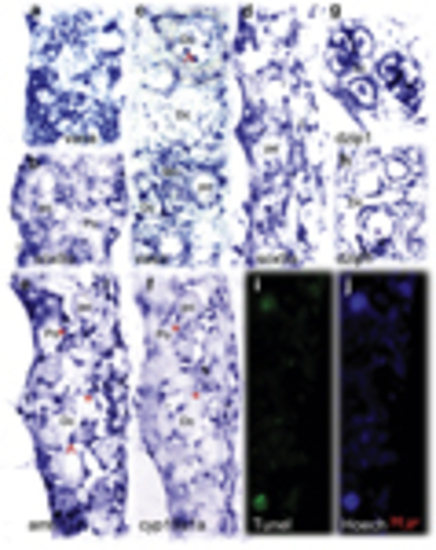
Comparative analysis of the expression pattern of multiple sex determinant genes between transforming and nontransforming gonads at 35 d.p.f. In the nontransforming gonads, vasa (a) was highly expressed whereas sox9a (b) expression was restricted in the two sides of the gonadal somatic cells. The transforming gonads revealed distinct expression patterns for vasa (c), sox9a (d), amh (e), cyp19a1a (f), and dzip1 (g and h). amh, cyp19a, and sox9a were expressed in somatic cells (supporting cells) located in the gonadal matrix and surrounding the oocytes. dzip1 was highly expressed in both spermatogonia and supporting cells. (e and f) Two adjacent sections with low levels of cyp19a1a and high levels of amh expressed with partial overlap in the same follicles (asterisk). Abnormally expressing aggregates in degenerative compartment (Dc) are indicated by the arrow. TUNEL staining assay showed the larger TUNEL-positive cells were consistent with degenerative oocytes (i). Hoechst staining (j) was used to count total number of gonadal cells. Go, giant oocyte; Mn, multinucleated giant cells; PO, perinucleolar oocyte. Scale bars=20μm |