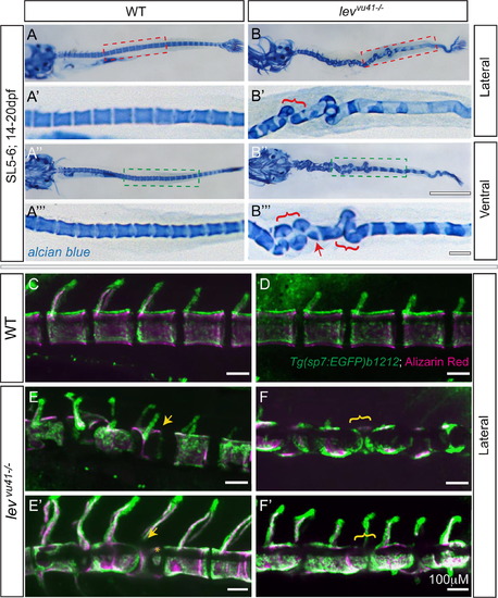
Segmentation of the vertebral column and localization of osteoblasts proceeds on a dysmorphic notochord template. Alcian blue stained larvae (14dpf) (A–B′′′) with lateral insets (red dashed box; A′, B′) and ventral insets (green dashed box; A′′′, B′′′). (A–A′′′) WT larvae display well-formed and segmented centra. (B–B′′′) levvu41-/- larvae display abberant apposition of centra (red brackets) and hemicentra (red arrow). Confocal projections of zebrafish vertebral column at 21dpf, illustrating boney matrix (alizarin red in magenta) and osteoblasts (Tg(sp7:EGFP)b1212 in green) (C–F′). (C, D) WT vertebrae and associated osteoblasts are well segmented along the vertebral axis. (E, E′) An individual levvu41-/- mutant displaying segmented colocalization of osteoblast and centra, including malformed hemivertebrae (yellow arrows) that continues to have mostly well segmented osteoblasts (E′), excepting some ectopic osteoblast-mineral formations in regions previously devoid of osteoblasts or mineral (E′, yellow asterix). (F and F′) An individual levvu41-/- mutant displaying regions of severe notochord bending with aberrantly apposed osteoblasts (F, yellow bracket), those progresses to a region with more complete osteoblast coverage (F′, yellow bracket). (C,D) 10dpf; ~SL3-4 (E,F) 10dpf; ~SL3-4 (E′, F′) 14dpf; ~SL4-5. Scale bars for (A, B, A3, B3)=1 mM; (C–F′) and (A′, A′′′,B′,B′′′)=100 mM.
|
