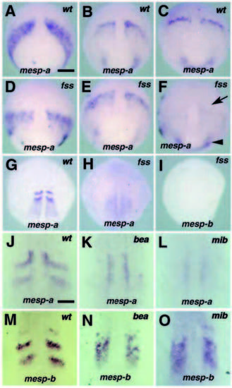
mesp expression in fss-type mutants. In all panels, the genotype of embryo is shown in the upper right corner and the probe used is at the bottom. (A-I) mesp expression in wild-type and fss embryos at 70% epiboly (A,D), 90% epiboly (B,E), 95% epiboly (C,F) and 12-somite stages (G-I). The normal expression pattern of mesp-a is seen in both wild-type and fss embryos up to 90% epiboly. In fss embryos, however, the striped expression is not maintained in later stages (arrow in F) while the expression at the blastoderm margin persists (arrowhead in F). Neither mesp-a nor mesp-b is expressed in fss mutants during segmentation (G-I). mesp expression in wild-type (J,M), bea (K,N) and mib (L,O) embryos at the 10- somite stage. mesp-b expression loses its striped pattern and shows a ‘salt and pepper’ pattern, which covers a region two- to three-somites wide in the paraxial mesoderm (N,O). As compared with mesp-b, mesp-a expression in the mutants is very weak and diffuse, and sometimes undetectable except for that in the adaxial cells (K,L). Bar in A, 100 μm; in J, 30 μm.
|
