
Fig. 2
- ID
- ZDB-FIG-140313-30
- Publication
- Takke et al., 1999 - her4, a zebrafish homologue of the Drosophila neurogenic gene E(spl), is a target of notch signalling
- Other Figures
- All Figure Page
- Back to All Figure Page
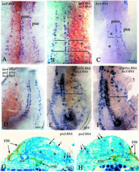
(A-F) Flat preparations of embryos labelled by in situ hybridization with an islet-1 probe (blue) and stained with an antibody against β-galactosidase (brown). pmn primary motoneurons, psn primary sensory neurons. Asterisks in all panels indicate the affected side. (A) 4-somite stage control embryo injected with lacZ RNA alone. (B-C) 4-somite stage embryos injected with full-length her4 and lacZ RNA. Both embryos have been stained for islet-1 expression, the one in B, in addition, for β-galactosidase expression. A reduction in the number of of islet-1-expressing cells can be observed on one side. The horizontal lines in these and the other panels indicate the extent of the enlargement of the neural plate on the affected side. (D) A 4-somite stage embryo injected with full-length her4, groucho2 and lacZ RNA, stained for islet-1 and β- galactosidase expression. Note the enlargement of the neural plate and the complete lack of islet-1-positive cells on the injected side. (E,F) Two 4-somite stage embryos which have been injected with deltaD(Pst) and lacZ RNA, and stained for her-4 and β-galactosidase expression; the anti-β-galactosidase staining was kept to a minimum in this case, to avoid brown overstaining. Notice that her-4 expression is reduced on the brown side. (G,H) Cross sections of 24 h embryos injected with groucho2 RNA and stained with two antibodies: F59, which recognises the myosin heavy chain (Miller et al., 1989) and labels the adaxial mesodermal cells and their derivatives (brown product; see Devoto et al., 1996), and Hu(C), a neuronal marker (Kim et al., 1996). Notice the asymmetry of the neural tube (not: notochord), one side of which is much larger. Hu(C) cells (blue product) are indicated with arrows on the affected side. Notice that mesodermal development, as judged by the F59 staining, is also affected on the same side. |