
Fig. 5
- ID
- ZDB-FIG-140306-3
- Publication
- Koshida et al., 1998 - Initial anteroposterior pattern of the zebrafish central nervous system is determined by differential competence of the epiblast
- Other Figures
- All Figure Page
- Back to All Figure Page
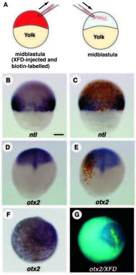
Role of FGF-R signalling in defining the posterior boundary of the otx2-expression domain. Embryos are oriented with the animal pole to the top. (A) Schematic representation of the experiment. The normal host blastula was transplanted with XFD-injected blastomeres. (B,C) The transplanted early gastrula (shield) stained with ntl probe (B), followed by biotin-peroxidase. The donor cells do not express ntl transcripts even in the marginal region. Note that the host marginal cells surrounded by XFD-donor cells tended to be less positive for ntl transcripts. This is probably due to the community effect (Gurdon et al., 1993) or the absorption of ligands by XFDoverexpressed donor cells. (D,E) The transplanted embryos (late gastrula, 10 h) were first stained with otx2 probe, followed by staining donor cells. Dorsal views of the same host before (D) and after (E) staining of the donor cells are shown. XFD-injected blastomeres (brown in E) strictly follow the host otx2-expression boundary. (F,G) The host embryo (late gastrula, 10 h) hybridized with DIG-labelled otx2 probe and fluorescein-labelled XFD probe is seen under transmitted light (F) or under ultraviolet light (G). The blastomeres containing XFD mRNAs (light green in G) follow the host otx2-expression boundary. Scale bar, 100 μm. |