
Fig. 1
- ID
- ZDB-FIG-140305-81
- Publication
- Koshida et al., 1998 - Initial anteroposterior pattern of the zebrafish central nervous system is determined by differential competence of the epiblast
- Other Figures
- All Figure Page
- Back to All Figure Page
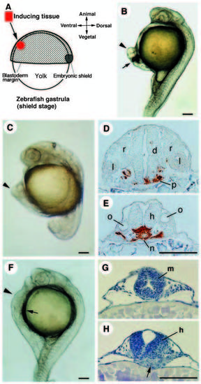
Secondary axes induced by transplantation of organizer tissues. Arrowheads in B, C and F indicate the secondary axis. (A) Schematic representation of organizer transplantation. A small piece of avian Hensen’s node (HN), fish organizer (embryonic shield) or Noggin/Chordin COS7 cell aggregate is transplanted into the host ventral region, mostly about halfway between the blastoderm margin and the animal pole, or near the margin in some cases. (B) Secondary axis induced by HN (stage 4) at 24 h. Arrow indicates the position of the transplanted HN. Chicken tissues appear dark because they contain yolk granules. (C-E) Secondary axis induced by the shield at 20 h. Cross sections at the level of the diencephalon (D) and the hindbrain (E) are shown. The donor cells are stained brown in the sections. Grafted shield differentiates into axial mesoderm and the ventral portion of the neural tube. (F-H) Secondary axis induced by Noggin/Chordin COS7 at 24 h. Cross sections at the level of the midbrain (G) and the hindbrain (H) are shown. Arrows indicate the position of the transplanted COS7 cells. COS7 cell mass is located under the induced neural tube (arrow in H). d, diencephalon; m, midbrain; h, hindbrain; l, lens; n, notochord; o, otic vesicle; p, prechordal plate; r, neural retina. Scale bars, 100 μm. |