
Fig. 5
- ID
- ZDB-FIG-140305-74
- Publication
- Appel et al., 1998 - Regulation of neuronal specification in the zebrafish spinal cord by Delta function
- Other Figures
- All Figure Page
- Back to All Figure Page
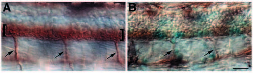
Disruption of lateral inhibition reduces the number of secondary motoneurons. (A) Lateral view of a 36 h control embryo labeled with zn5, which identifies secondary motoneurons (brackets) and ventral nerves (arrows) exiting the spinal cord. (B) Similar view of a 36 h embryo injected with X-Delta-1STU and nlacZ mRNAs. Lineage tracer (blue staining) is expressed in small clusters of spinal cord cells. Outside the clusters, zn5 labeling looks essentially normal. Cells within lineage-marked clusters are largely zn5-negative, showing that secondary motoneurons have not differentiated. Ventral nerves (arrows) appear reduced in size, suggesting the presence of fewer motor axons than in normal embryos. Scale bar, 20 μM. |