
Fig. 3
- ID
- ZDB-FIG-140305-71
- Publication
- Appel et al., 1998 - Regulation of neuronal specification in the zebrafish spinal cord by Delta function
- Other Figures
- All Figure Page
- Back to All Figure Page
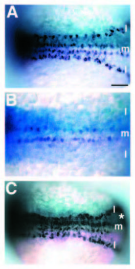
Delta function regulates neurogenesis. Dorsal views (anterior left) of 6-somite-stage embryos injected with mRNA at early blastula stages and probed for huC expression. In each case, b-galactosidase activity, as lineage tracer, is revealed as light blue staining and huC expression as dark blue. (A) Embryo injected with nlacZ mRNA only. huC is expressed in discontinuous, bilateral rows of cells in medial (m) and lateral (l) regions of neural keel. (B) Embryo injected with full-length deltaA and nlacZ mRNAs. Lineage tracer is distributed thoughout the embryo, with the right side (upper portion) receiving more than the left (lower). huC expression is absent from both lateral domains and the number of huC-expressing cells in the medial rows is reduced, particularly on the right side. Lateral domains seem more sensitive than medial domains to increased deltaA expression (two independent experiments: 40% and 47% of embryos with decreased medial huC expression while greater than 90% of embryos show reduced lateral huC expression). (C) Embryo co-injected with nlacZ and X-Delta-1STU mRNA. Lineage tracer shows that the right side (asterisk) received most of the injected mRNA. The density of huC-labeled neurons is increased in medial and lateral regions of neural keel receiving injected mRNA. Scale bar, 50 μm. |