
Fig. 3
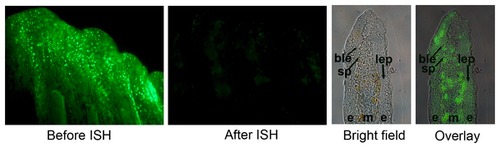
Morpholinos target all cellular compartments of the regenerating fin. Fluorescein-positive cells successfully took up the morpholino (1 day post electroporation). The two left panels reveal loss of fluorescein signal following in situ hybridization. The two right panels demonstrate that fluorescein-positive cells are observed in all cellular compartments in freshly harvested and cryosectioned fins. The basal layer of the epidermis (ble) is identified as a row of cuboidal cells between the epithelium (e) and the mesenchyme (m). The skeletal precursor cells (sp) are located adjacent to the ble. Morpholino uptake is observed in the outer epithelial layers, in the skeletal precursors, and in the medial mesenchyme. |