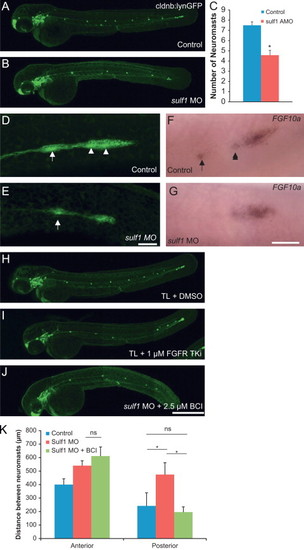
sulf1 knockdown leads to loss of FGF signaling within the lateral line primordium but can be partially rescued by BCI. (A) Whole mount Tg(cldnb:lyngfp) zebrafish at 54 hpf showing normal distribution of posterior lateral line (pLL) neuromasts. (B) Whole mount Tg(cldnb:lynGFP) sulf1 morphant zebrafish at 54 hpf showing a decreased number of pLL neuromasts along the tail, quantified in the adjacent graph. (C) Quantification of neuromast number from control and sulf1 morphant zebrafish. Morphants have 4.6±0.6 neuromasts while controls have 7.5±0.3 neuromasts along their trunk. (D) View of the pLL primordium from a control Tg(cldnb:lynGFP) zebrafish at ∼32 hpf shortly after deposition of a neuromast (arrow), where the rosette patterning of the next two neuromasts to be deposited can be seen (arrows). (E) View of the pLL primordium of a Tg(cldnb:lynGFP) sulf1 morphant zebrafish at ∼32 hpf shortly after deposition of a neuromast (arrow). The primordium is much smaller than in controls, and the typical rosette patterns within the neuromast or in the trailing edge of the primordium are not visible. (F and G) In situ hybridization of an FGF10a probe in control (F) or sulf1 morphant (G) zebrafish at 36 hpf. FGF10a is expressed in the leading zone of the primordium, and in the central region of a recently deposited neuromast (arrow) and at the center of the rosette of the next neuromast to be deposited (arrowhead). In sulf1 morphants, FGF10a is expressed more uniformly across the smaller neuromast and is not focally expressed at the center of a rosette along the trailing edge. (H and I) Tg(cldnb:lynGFP) zebrafish treated at 24 hpf with either DMSO (H) or 1 μM FGFRTKi (I) imaged at 54 hpf showing that partial inhibition of FGFR1 can phenocopy the reduced number of deposited neuromasts. (J) sulf1 morphant Tg(cldnb:lynGFP) zebrafish treated at 32 hpf with 2.5 μM BCI and imaged at 54 hpf, showing a decrease in the spacing between the last 4 deposited neuromasts compared with the initial two. (K) Quantification of the distance between the first two deposited neuromasts (anterior) and last two deposited neuromasts excluding the terminal cluster (posterior) at 54 hpf from control fish (blue), sulf1 morphant fish treated with DMSO (red), and sulf1 morphant fish treated at 32 hpf with 2.5 μM BCI (green). BCI had no effect on neuromast distance in the anterior neuromasts that had already been deposited before it was added (p>0.05 compared to DMSO-treated morphants; ANOVA), but restored the appropriate distance between posterior neuromasts deposited after its application (p<0.001 compared to DMSO-treated morphants; p>0.05 compared to control; ANOVA). Scale bars: A, B, H, I, J: 500 μm; D, E: 50 μm; F, G: 50 μm.
|
