
Fig. 3
- ID
- ZDB-FIG-140122-41
- Publication
- Kang et al., 2013 - Local dkk1 crosstalk from breeding ornaments impedes regeneration of injured male zebrafish fins
- Other Figures
- All Figure Page
- Back to All Figure Page
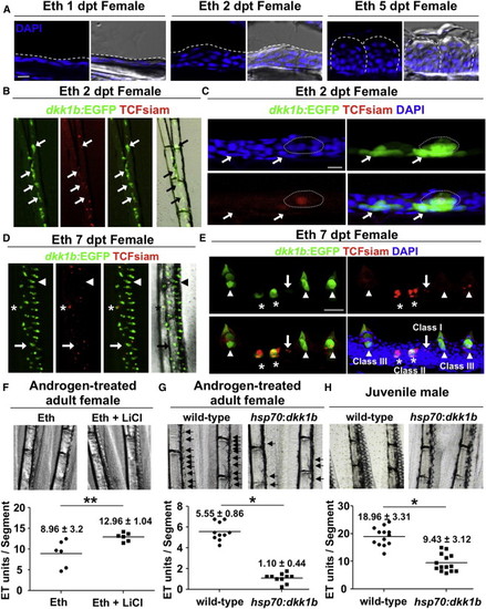
Induction of De Novo Formation of ETs by Androgen and Wnt Signaling (A) Section images of the dorsal side of a female pectoral fin after the indicated duration of androgen treatment. Dashed lines indicate the outer epidermal border (left and middle images) or individual ETs (right images). Note that local epidermal thickening is observed by 2 dpt. Scale bar, 10 μm. (B and C) Whole-mount (B) and section (C) images of female TCFsiam; dkk1b:EGFP pectoral fins after 2 days of androgen treatment. Arrows indicate dkk1b:EGFP expression in basal layers in forming ETs. Dotted line outlines an ET precursor, with one middle cell expressing weak TCFsiam. Scale bar in (C), 10 μm. (D and E) Whole-mount (D) and section (E) images of female TCFsiam;dkk1b:EGFP pectoral fins after 7 days of androgen treatment. ETs show stage-specific expression profiles. Putative newly forming ETs express TCFsiam (Class I, arrow). Putative immature ETs show both TCFsiam and dkk1b:EGFP expressions (Class II, asterisks). Mature ETs express only dkk1b:EGFP or both TCFsiam and dkk1b:EGFP (Class III, arrowheads). Note that TCFsiam expression is weak in mature ETs. Scale bar in (E), 50 μm. (F) LiCl treatment increases formation of ETs in androgen-treated adult females. Data are mean ± SD. p < 0.05 by two-tailed Student’s t test. (G and H) Ubiquitous overexpression of dkk1b blunts ET formation in androgen-treated adult females (G) and juvenile males (H). Arrows indicate developing ETs in (G). Data are mean ± SD. p < 0.001 by two-tailed Student’s t test. |
Reprinted from Developmental Cell, 27(1), Kang, J., Nachtrab, G., and Poss, K.D., Local dkk1 crosstalk from breeding ornaments impedes regeneration of injured male zebrafish fins, 19-31, Copyright (2013) with permission from Elsevier. Full text @ Dev. Cell