
Fig. 1
- ID
- ZDB-FIG-140122-2
- Publication
- Hayes et al., 2013 - Spinal Deformity in Aged Zebrafish Is Accompanied by Degenerative Changes to Their Vertebrae that Resemble Osteoarthritis
- Other Figures
- All Figure Page
- Back to All Figure Page
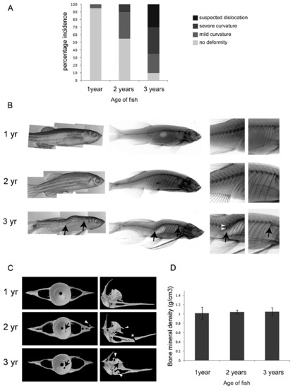
Aging zebrafish show gross morphological changes to the vertebral column. A. Graph showing incidence (%) of deformities by age. n=20 for each age group. B. Gross morphological appearance (left panel) and corresponding radiology (middle and left panels; left panels show detail of trunk and tail vertebrae) of zebrafish at 1, 2 and 3-years. Black arrows (in bottom panel) denote suspected dislocations of the spine. White arrowheads point to regions of increased bone density in vertebrae surrounding the dislocation. C. MicroCT images showing a representative single reconstructed vertebra (C5) from each age group, black arrows point to regions of bone erosion, white arrowheads point to bony outgrowths; asterisk denotes fracture. D. Graph of average bone mineral density shows no difference to bone density at the different ages, tested by One-way ANOVA; 1 vs 2 year P=0.80, 1 vs 3 year P=0.92, 2 vs 3 year P=0.79. n=3 for each age. |