
FIGURE
Fig. S4
- ID
- ZDB-FIG-140114-62
- Publication
- Leigh et al., 2013 - Mmp17b is essential for proper neural crest cell migration in vivo
- Other Figures
- All Figure Page
- Back to All Figure Page
Fig. S4
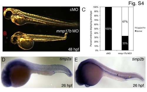
Cartilage and vascular defects observed in mmp17b knockdown fish. A-B are bright field images of control MO (A) and mmp17b MO1-injected (B) 48 hpf embryos. Panel B illustrates the cupped-fin phenotype seen in mmp17b MO1-injected embryos compared to controls. This defect was quantitated in panel C that shows mmp17b MO1-injected embryos have a statistically significant increase in the cupped-fin defect compared to controls. Panels D and E show timp2a and timp2b 26 hpf ISH embryos (head is to the left). Yellow arrow in D shows plexus region and the black arrow indicates regions adjoining the vasculature. |
Expression Data
| Genes: | |
|---|---|
| Fish: | |
| Anatomical Terms: | |
| Stage: | Prim-5 |
Expression Detail
Antibody Labeling
Phenotype Data
| Fish: | |
|---|---|
| Knockdown Reagent: | |
| Observed In: | |
| Stage: | Long-pec |
Phenotype Detail
Acknowledgments
This image is the copyrighted work of the attributed author or publisher, and
ZFIN has permission only to display this image to its users.
Additional permissions should be obtained from the applicable author or publisher of the image.
Full text @ PLoS One