|
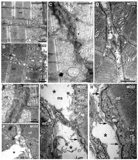
TEM reveals defects in MTJ and myosepta ultrastucture, and identifies the rupture site at the outer surface of the basement membrane. Micrographs of 5 dpf uninjected (A,C,E) and MO22-injected (B,D,F-H) larvae. (A,B) Sarcomeric organization of skeletal muscle in morphants (B) and uninjected larvae (A). (C,D) Detail of the sarcolemma and basement membrane structure (arrows) in wild type (C) and morphants (D). (E-H) Detail of MTJ and myoseptum structure in wild type (E) and morphants (F-H). Asterisks show a gap corresponding to detached and retracted fiber (F) and tears between myoseptum and muscle fibers (G,H). At rupture sites, basement membrane remains attached to sarcolemma (G,H, arrows) and is torn away with fiber detachment and retraction (F, arrow). Contrary to control (E), fibroblasts (fb) are observed in damaged myosepta (F-H). ms, myosepta; mf, muscle fiber.
|