
FIGURE
Fig. S2
- ID
- ZDB-FIG-131218-20
- Publication
- Sittaramane et al., 2013 - The PCP protein Vangl2 regulates migration of hindbrain motor neurons by acting in floor plate cells, and independently of cilia function
- Other Figures
- All Figure Page
- Back to All Figure Page
Fig. S2
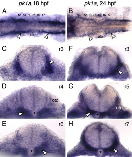
pk1a is expressed primarily outside the neural tube in the paraxial mesoderm and endoderm. (A,B) Dorsal views of zebrafish hindbrains at 18 and 24 hpf processed for pk1a in situs are suggestive of pk1a expression in the lateral regions of the neural tube (arrowheads). However, cross-sections at various levels through the hindbrain (C-H) reveal that pk1a is expressed primarily in the paraxial mesoderm and endoderm (arrowheads). Asterisks and oto indicate notochord and otic vesicle, respectively. |
Expression Data
Expression Detail
Antibody Labeling
Phenotype Data
Phenotype Detail
Acknowledgments
This image is the copyrighted work of the attributed author or publisher, and
ZFIN has permission only to display this image to its users.
Additional permissions should be obtained from the applicable author or publisher of the image.
Reprinted from Developmental Biology, 382(2), Sittaramane, V., Pan, X., Glasco, D.M., Huang, P., Gurung, S., Bock, A., Li, S., Wang, H., Kawakami, K., Matise, M.P., and Chandrasekhar, A., The PCP protein Vangl2 regulates migration of hindbrain motor neurons by acting in floor plate cells, and independently of cilia function, 400-412, Copyright (2013) with permission from Elsevier. Full text @ Dev. Biol.