
Fig. 1
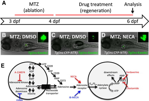
A Chemical Screen for β Cell Regeneration (A) Schema for the β cell regeneration screen. The pancreatic β cells are conditionally targeted for ablation from 3-4 dpf by using β cell-specific expression of nitroreductase (Tg[ins:CFP-NTR]), which converts metronidazole (MTZ) into a cytotoxic product. After washing away the MTZ, the larvae are placed in 96-well plates and exposed to 2–50 μM of the compounds in 1% DMSO. After 2 days of recovery, from 4-6 dpf, β cell regeneration can be easily quantified in double-transgenic larvae, Tg(ins:CFP-NTR);Tg(ins:Kaede), because Kaede labels the β cells with bright fluorescence. (B) Picture of a control Tg(ins:Kaede) larva at 6 dpf that was not affected by the MTZ treatment (because it does not express NTR) and therefore displays a typical number of β cells, as visualized with the microscope used for screening. The inset displays a magnified view of the pancreatic islet (outlined by the dashed square). (C) Tg(ins:CFP-NTR);Tg(ins:Kaede) larva (6 dpf) following β cell ablation with MTZ from 3-4 dpf and vehicle treatment from 4-6 dpf. Typically, these control larvae have three to seven β cells at this stage. (D) Tg(ins:CFP-NTR);Tg(ins:Kaede) larva following β cell ablation with MTZ from 3-4 dpf and treatment with the hit compound NECA from 4-6 dpf. This particular larva contains too many β cells to count without the use of confocal microscopy. (E) The hit compounds converge on adenosine signaling/metabolism by targeting adenosine kinase (A-134974), adenosine receptors (NECA), and phosphodiesterase 3/4 (Cilostamide and Zardaverine). Compounds that increased β cell regeneration more than 2-fold after 2 days of treatment are labeled in red. After rescreening all activators of adenosine signaling contained in the Sigma LOPAC library, we found that an adenosine deaminase inhibitor (EHNA) and another nonselective adenosine agonist (IB-MECA) (labeled in blue) could also increase β cell regeneration, although less than 2-fold. See also Figure S1. |
| Gene: | |
|---|---|
| Fish: | |
| Conditions: | |
| Anatomical Term: | |
| Stage: | Day 6 |
| Fish: | |
|---|---|
| Conditions: | |
| Observed In: | |
| Stage: | Day 6 |
Reprinted from Cell Metabolism, 15(6), Andersson, O., Adams, B.A., Yoo, D., Ellis, G.C., Gut, P., Anderson, R.M., German, M.S., and Stainier, D.Y., Adenosine Signaling Promotes Regeneration of Pancreatic beta Cells In Vivo, 885-894, Copyright (2012) with permission from Elsevier. Full text @ Cell Metab.