
Fig. 2
- ID
- ZDB-FIG-130912-27
- Publication
- Sandoval et al., 2013 - Juxtaposition of chemical and mutation-induced developmental defects in zebrafish reveal a copper-chelating activity for kalihinol f
- Other Figures
- All Figure Page
- Back to All Figure Page
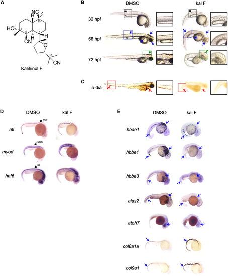
Embryos Treated with Kalihinol F Show a Phenotype Consistent with Copper Deficiency (A) Chemical structure of kalihinol F. (B) Exposure of zebrafish embryos to 2.5 μg/ml of kalihinol F (kal F) resulted in a wavy notochord (black arrow, D, ntl), loss of pigmentation (blue arrow) and enlarged hindbrain vesicle (green arrow) as compared to DMSO-treated control embryos. Boxed figures are enlargements of highlighted portions on whole embryos. (C) o-dianisidine staining at 72 hpf revealed loss of hematopoiesis (red arrows) in treated embryos versus control. (D) In situ hybridization on 32 hpf embryos for ntl, myod, and hnf6. not, notochord; som, somites; and sc, spinal cord. (E) In situ hybridization for hbae1, hbbe1, hbbe3, alas2, atoh7, col8a1a, and col9a1 confirms gene expression analysis by RNA sequencing that is consistent with copper-deficient phenotype of kalihinol F. See also Tables S1 and S2. |