
Fig. S1
- ID
- ZDB-FIG-130828-7
- Publication
- Sacilotto et al., 2013 - Analysis of Dll4 regulation reveals a combinatorial role for Sox and Notch in arterial development
- Other Figures
- All Figure Page
- Back to All Figure Page
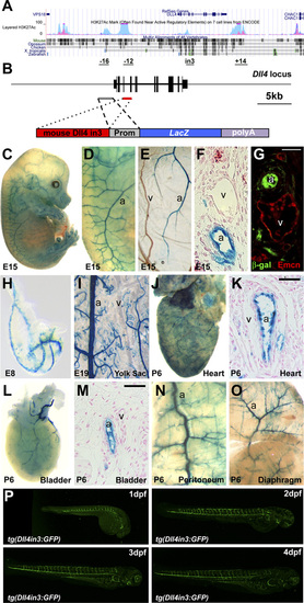
(A) Schematic representation of the Dll4 locus from University of California, Santa Cruz (UCSC) ENCODE Browser (1), HUVEC-specific H3K27Ac in light blue, K562 leukemia cell line in pink. On the bottom, the potential vascular-specific enhancers are depicted with their respective positions (in kb) with respect to the TSS (except for the intron 3, indicated as in3). (B) Schematic representation of the mouse Dll4 locus (Upper line, exons are black boxes) and Dll4in3end transgene (Lower line). Prom, endogenous promoter. (C–G) A representative X-Gal–stained E15 Dll4in3end transgenic embryo. X-Gal staining is detected in arterial (a) but not venous (v) cells in whole-mount embryo (C and D), yolk sac (E), and transverse section (F). Expression of the venous marker Endomucin (Emcn) did not overlap that of the reporter gene (G). (H–O) Further examples of transgenic mice expressing the mouse Dll4in3hsp transgene at E8 (H), E19 (I), and tissues from a P6 pup (J–O), all showing X-Gal staining in arteries (a) but not veins (v). (P) Time course of the tg(Dll4in3:GFP) zebrafish line between 1–4 dpf. |