
Fig. S1
- ID
- ZDB-FIG-130808-57
- Publication
- Lee et al., 2013 - An exclusively mesodermal origin of fin mesenchyme demonstrates that zebrafish trunk neural crest does not generate ectomesenchyme
- Other Figures
- All Figure Page
- Back to All Figure Page
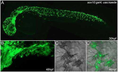
Neural crest cells are well labelled in sox10:gal4; uas:kaede embryos. Lateral confocal images of 30-hpf (A,C,C′) and 48-hpf (B) sox10:gal4; uas:kaede transgenic embryos immunostained with an antibody detecting Kaede. (A) Broad and robust neural crest labelling along the entire axis can be seen at 30 hpf. (B) Lateral view of the branchial arches at 48 hpf showing extensive labelling of presumptive ectomesenchymal neural crest. (C,C′) Images showing DIC alone (C) or overlaid on a fluorescent image of Kaede expression demonstrates labelling of melanophores in sox10:gal4; uas:kaede transgenic embryos (C′). In this image, it can be seen that all five melanocytes express Kaede. Quantification of five 30-hpf sox10:gal4; uas:kaede transgenic embryos showed that at least 93.0% of melanophores are labelled by Kaede (a total of 545 melanophores was counted). |