
Fig. 6
- ID
- ZDB-FIG-130808-56
- Publication
- Lee et al., 2013 - An exclusively mesodermal origin of fin mesenchyme demonstrates that zebrafish trunk neural crest does not generate ectomesenchyme
- Other Figures
- All Figure Page
- Back to All Figure Page
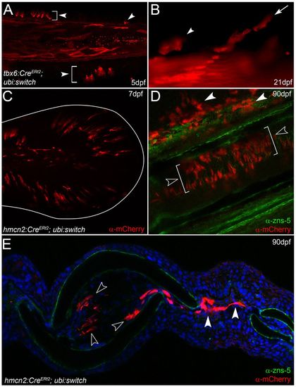
Fin mesenchyme contributes to the adult fin fibroblasts. (A,B) tbx6:CreERt2; ubi:switch transgenics treated with 4-hydroxytamoxifen and imaged at 5 dpf (A) and 21 dpf (B). Arrowheads indicate fin mesenchyme cells in the larval fin (A) and retained in juvenile fin (B). Chains of cells are seen invading at 21 dpf (arrow in B). (C-E) Images of ubi:switch transgenics injected with hmcn2:CreERt2 BAC transgene and treated with 4-hydroxytamoxifen from 3-4 dpf. (C) Tail region of 7-dpf larva shows mCherry in fin mesenchyme cells. The extent of the fin is outlined. (D,E) Adult fins immunostained for mCherry (red) and with zns-5 antibody (green) imaged in lateral whole-mount (D) or in transverse view following cryosectioning and counterstaining with DAPI (E). mCherry cells are in locations consistent with fibroblasts and are zns-5 negative. They reside within the fin rays (open arrowheads in D,E) or can be seen in the inter-ray region (white arrowheads in D,E). |
| Gene: | |
|---|---|
| Fish: | |
| Condition: | |
| Anatomical Term: | |
| Stage Range: | Day 5 to Days 21-29 |