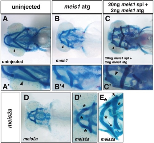
Cartilage rotation and fusion defects in meis1 and meis2a morphants. A, Ventral view of viscerocranial cartilages of a 5dpf uninjected larva. A′, Magnified view of A showing position of palatoquadrate (arrowhead). B, Ventral view of viscerocranial cartilages of a 5dpf meis1 ATG morphant larva. B′, Magnified view of B showing cartilage fusions in morphants as well as an apparent rotation of the palatoquadrate cartilage (arrowhead). C, Ventral view of viscerocranial cartilages of a 5pf morphant larva co-injected with both meis1 ATG and meis1 SPL MOs. C′, Magnified view of C showing cartilage fusions in morphants and the apparent rotation of the palatoquadrate cartilage (arrowhead). D, Ventral view of viscerocranial cartilages of a 5dpf meis2a ATG morphant larva. D′, Magnified view of D showing cartilage fusions (asterisks) in morphants. E, Ventral view of viscerocranial cartilages that were dissected and flat-mounted from a 5dpf meis2a ATG morphant larva showing cartilage fusions (asterisks). Arrow indicates where a cartilage fusion was broken during dissection.
|
