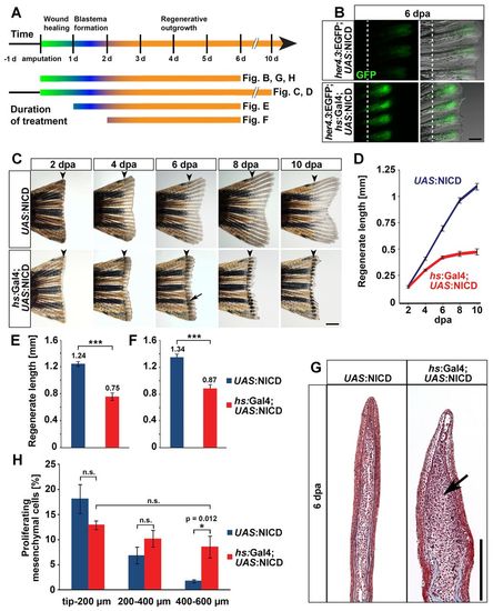
NICD overexpression expands the proliferative blastema, yet stalls fin regeneration. (A) Schematic timeline of fin regeneration and experimental treatments. (B) Enhancement of EGFP expression in the blastema of her4.3:EGFP; hs:Gal4; UAS:NICD triple transgenic fins at 6 dpa after repeated heatshocks starting at the time of amputation. Dashed line indicates amputation plane. Scale bar: 200 μm. (C) Inhibition of regeneration in hs:Gal4; UAS:NICD fish heatshocked repeatedly starting 1 day prior to fin amputation. Pigment cells cluster in the stalled regenerate (arrow). Arrowheads indicate amputation plane. Scale bar: 500 μm. (D) Regenerate lengths of fins treated as in C; mean±s.e.m. Length differences at all timepoints, except 2 dpa, are highly significant (P<0.001). UAS:NICD: n=8 fins, 24 fin rays; hs:Gal4; UAS:NICD: n=12 fins, 36 fin rays. (E,F) NICD overexpression starting at 1 dpa (E) or 2 dpa (F) is able to delay fin regeneration, mean±s.e.m. n=8 fins, 24 fin rays each group. ***P<0.001. (G) Masson’s trichrome staining reveals expansion of the mesenchymal compartment of regenerates (arrow) after sustained NICD overexpression for 6 days. Scale bar: 200 μm. (H) Percentage of BrdU-positive mesenchymal cells in three 200 μm regions along the distal-proximal axis of UAS:NICD and hs:Gal4; UAS:NICD fins heatshocked repeatedly for 6 days. NICD overexpression increases proliferation in the scarcely proliferating proximal 400-600 μm domain (‘differentiation zone’). n=5 fish, six sections. *P<0.05.
|
