
Fig. 4
- ID
- ZDB-FIG-130618-19
- Publication
- Villefranc et al., 2013 - A truncation allele in vascular endothelial growth factor c reveals distinct modes of signaling during lymphatic and vascular development
- Other Figures
- All Figure Page
- Back to All Figure Page
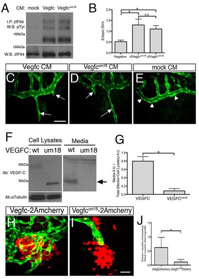
um18 mutation affects Vegfc secretion. (A) Western blot (W.B.) of Flt4 immunoprecipitates (I.P.) from cells treated with the indicated conditioned medium (CM), probed with antibodies against phosphotyrosine (pTyr) and zebrafish Flt4. (B) Quantification of ectopic subintestinal vessels (SIVs) in embryos injected with the indicated CM. Values are the average of three experiments. (C-E) Confocal micrographs of SIVs (arrowheads) in Tg(fli1a:egfp)y1 embryos at 3.5 dpf following perivitelline injections with (C) Vegfc CM, (D) Vegfcum18 CM or (E) mock CM. Arrows indicate ectopic SIVs. (F) Western blots probed with the indicated antibodies. Cell lysates and media from NIH3T3 cells transiently expressing human VEGFC or VEGFCum18. Arrow indicates processed secreted form of wild-type VEGFC. (G) Quantitation of western blots in F. Values are the average of three experiments. A.U., arbitrary units. (H,I). Confocal image of xenografts in 4.5-dpf Tg(fli1a:egfp)y1 embryos expressing (H) Vegfc-P2Amcherry or (I) Vegfcum18-P2Amcherry (pseudocolored red). (J) Quantification of vascular density in xenografts from three experiments. (C-E,H,I) Anterior is to the left, dorsal is up. *P<0.05; N.S., not significant; error bars indicate s.e.m. Scale bars: 50 μm in C-E; 10 μm in H,I. |