
Fig. 6
- ID
- ZDB-FIG-130530-2
- Publication
- Fernandez et al., 2013 - Fixation/permeabilization: New alternative procedure for immunofluorescence and mRNA in situ hybridization of vertebrate and invertebrate embryos
- Other Figures
- All Figure Page
- Back to All Figure Page
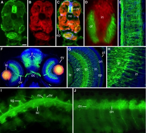
Immunofluorescent staining of freeze-sectioned 3-day-old (A–H) and whole-mounted 6-day-old (I,J) zebrafish larvae. A–C: Microtubule-stained (A), actin-stained (B), and merged image (C) of a triple stained (DAPI) larva. Microtubules and actin filaments co-distribute in the outer sector of the neural rod (nr) and developing musculature (mu). ep, epidermis; no, empty notochord [Form-Acet]. D: Double-stained (microtubules, green; nuclei, red) merge image of the neural rod showing its inner nuclear (in) and outer fibrillar (of) sectors [Form-Acet]. E: Confocal deconvolved double-stained image (microtubules green, nuclei blue) of a longitudinally sectioned neural rod [Form-Acet]. F: Triple-stained (microtubules, green; β-catenin, red; nuclei, blue) merged image of a coronal section along the head of a larva showing the eye (ey), optic nerve (ov), optic chiasm (oc), and tectal fiber tracts (tf). The central lens (le) cells express β-catenin and microtubules [Form-Acet]. G: Confocal deconvolved merge image of a double-stained (microtubules, green; nuclei, blue) eye showing the retina layers: pt, photoreceptors; on/op, outer nuclear and plexiform layers; in/ip, inner nuclear and plexiform layers; gc, ganglion cell layer; le, lens [Form-Acet]. H: Sector of the same retina showing the inner nuclear (in) and plexiform (ip) layers. I: Spinal cord (sc), nerve roots (nv), and sensory ganglia (sg) stained for microtubules. J: Optical section of a similarly stained spinal cord with developing neurons (dn) [Form-Acet]. Scale bar = 25 μm in A–C, 10 μm in D,E, 35 μm in F, 15 μm in G, 6 μm in H, 25 μm in I,J. |