
Fig. 5
- ID
- ZDB-FIG-130530-1
- Publication
- Fernandez et al., 2013 - Fixation/permeabilization: New alternative procedure for immunofluorescence and mRNA in situ hybridization of vertebrate and invertebrate embryos
- Other Figures
- All Figure Page
- Back to All Figure Page
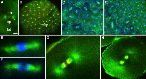
Immunofluorescence staining of zebrafish whole-mounted embryos. A: Double stained (microtubules green, chromosomes red) metaphase first cleavage spindle (sp). The arrow points to the DNA left by the extruded second pole cell. bd, blastodisc [Form-Acet-Peg]. B: Similarly stained blastula with numerous methaphase mitotic spindles (sp) [Form-Acet]. C,D: Confocal images of advanced blastulae showing asynchronously dividing blastodermal cells (microtubules, green; nuclei, blue) prepared with our method [Form-Acet] and stained for β-tubulin (C) or according to Solnica-Krezel and Driever (1994) (D). E,F: High magnification double-stained (microtubules, green; chromosomes, blue) metaphase (E) and anaphase (F) mitotic spindles stained for tyrosinated tubulin [Form-Acet-Peg]. G,H: Confocal images (microtubules, green; chromosomes, red) of anaphase mitotic spindles stained for tyrosinated (G) and α-tubulin (H). The latter image was deconvolved. Notice the successful staining of peripheral and astral microtubules [nForm-Acet-DMSO-Tx]. Scale bar = 30 μm in A, 55 μm in B, 25 μm in C,D, 3 μm in E,F, 10 μm in G,H. |