|
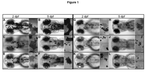
Dorsal views from five insertional zebrafish mutants in V-ATPase subunits. The fish larvae are shown at 2 dpf (A, C, E, G, I and K) and at 5 dpf (B, D, F, H, J and L). For each image a close up view is at the right side. Wild type larvae are in (A and B) and show normal pigmentation. Pigment dilution is observed in all V-ATPase mutants (C – L), however the severity of the pigment phenotype was mild in V0-d1 (gene atp6vod1) (C and D) and V0-ac45b (gene atp6ap1b) (E and F). It was more severe in V1-E1b (gene atp6v1e1b) (G and H) and V1-H (gene atp6v1h) (I and J), but even more severe in V0-ca (gene atp6v0ca) (K and L). In all the mutants there are a combination of pale melanocytes and dark melanin spots considered to be fragments of cells. Pale melanocytes became scarce the more severe the phenotype was (compare C and I). Bars in low amplification views are 200 μm and bars in close ups are 20 μm.
|