|
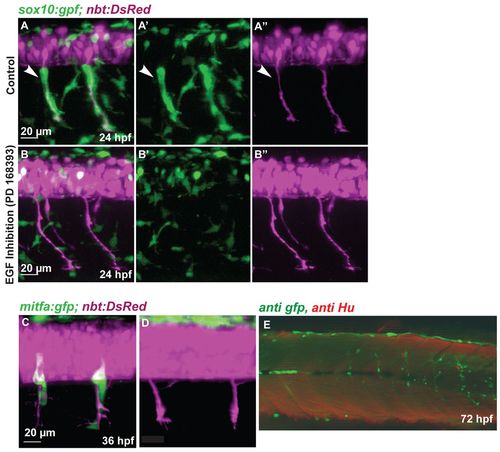
NC migration along the ventromedial path is blocked by inhibition of ErbB receptors. (A-B′′) Tg(sox10:gfp; nbt:DsRed) zebrafish embryos at 24 hpf. (A,B) Red and green channel (merge); (A′,B′) green channel; (A′′,B′′) red channel. Medial NC cells (green) covering the primary motor axons (white arrowheads, A-A′′) are absent after treatment with the ErbB inhibitor PD168393 (B-B′′). (C,D) Confocal images of 36 hpf Tg(mitfa:gfp; nbt:DsRed) embryos. (C) Wild-type embryo. (D) Embryo treated with ErbB inhibitor at 16 hpf. (E) Tg(mitfa:gfp) embryos were injected with a double MO combination against mitfa and erbb3b. Larvae were stained at 8 dpf using anti-GFP (green) and anti-HU (red) antibody (white arrowheads). The association of DRGs (red) and GFP-positive cells was quantified (Table 1). In double mitfa and erbb3b knockdowns, of 82 metamers lacking HU positive cells only five (6%) develop a string of GFP-positive cells.
|