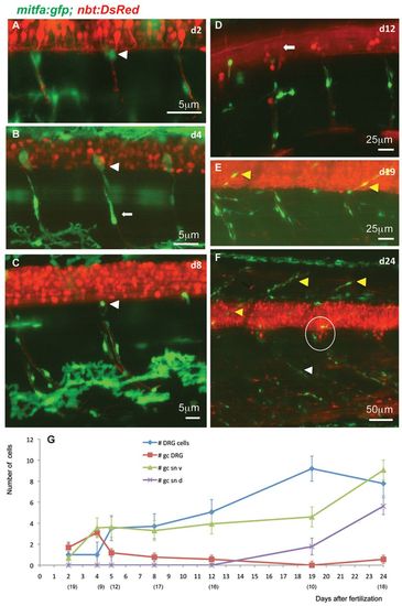
Stationary MPs located at the DRGs give rise to MPs located along the spinal nerves. (A-F) Time-lapse confocal imaging of Tg(mitfa:gfp;nbt:DsRed) zebrafish individuals during larval development of consecutive ages. GFP-positive cells at the exit point of the spinal nerves (white arrowheads in A-C) appear to be stationary. Other GFP-positive cells of more elongated shapes are observed along the spinal nerves (e.g. arrow in B). Until 12 dpf (D), no GFP-positive cells can be seen along the dorsally extending spinal nerves (arrow in D), whereas at metamorphic stages (E,F) both the dorsally extending (yellow arrowheads) and the ventrally extending (white arrowhead) spinal nerves show an increased number of associated GFP-positive cells. The number of cells in each DRG (circled in F) has increased to about eight. (G) Average numbers of GFP-positive cells per hemisegment located at the DRGs, the ventral and dorsal spinal nerves as well as DRGs at increasing larval age (dpf). The numbers of hemisegments counted is indicated below the respective time points. gc DRG, green cells at DRG; gc sn v, green cells at spinal nerves ventral; gc sn d, green cells at spinal nerves dorsal.
|
