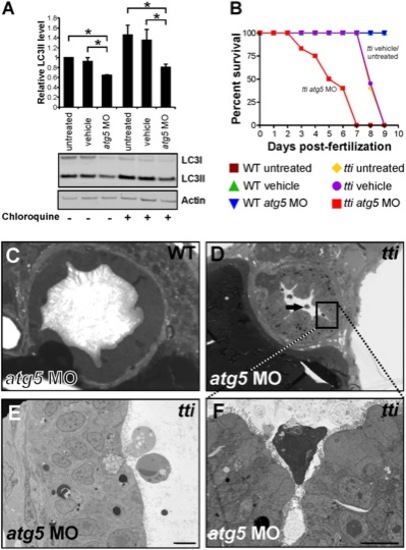
(A) Western blot analysis of lysates of ttis450 larvae (72 hpf) that had been injected at the 1–4 cell stage with an antisense morpholino oligonucleotide (MO) targeted to the start codon of atg5 mRNA reveals decreased levels of LC3II compared to untreated and vehicle controls, both in the presence and absence of chloroquine. Data are represented as mean +/ SD, *p<0.05. (B) Survival curve of untreated WT and ttis450 larvae compared to WT and ttis450 larvae that had been injected at the 1–4 cell stage with vehicle or atg5 MO (n>85 larvae per group). The lifespan of WT embryos/larvae is completely unaffected by injection with the atg5 MO since all three groups of WT larvae (untreated, vehicle-treated and atg5 MO-treated) progress normally through the first 10 days of development, when the experiment was terminated. The horizontal line represents untreated WT embryos (maroon squares), vehicle-injected WT embryos (green triangles) and atg5 MO-injected WT embryos (blue triangles). In contrast, ttis450 embryos respond to microinjection of the atg5 MO by impaired survival. Whereas all untreated (yellow diamonds) or vehicle-injected (purple circles) ttis450 larvae are still alive at 7 dpf, all the atg5 MO-injected ttis450 larvae are dead at this time-point (red squares). Indeed, 20% of the atg5 MO-injected ttis450 larvae have already succumbed by 3 dpf. (C–F) TEMs of WT (C) and ttis450 larvae at 120 hpf (D–F), injected at the 1–4 cell stage with the atg5-targeted MO. Inhibiting autophagy in ttis450 larvae results in the appearance of detached and shrunken IECs in the intestinal lumen (black arrow in D, E and F [boxed area in D]) but has no impact on WT IECs (C). Scale bars = 10 μm.
|
