
FIGURE
Fig. 4
- ID
- ZDB-FIG-130404-28
- Publication
- Cannon et al., 2013 - Global analysis of the haematopoietic and endothelial transcriptome during zebrafish development
- Other Figures
- All Figure Page
- Back to All Figure Page
Fig. 4
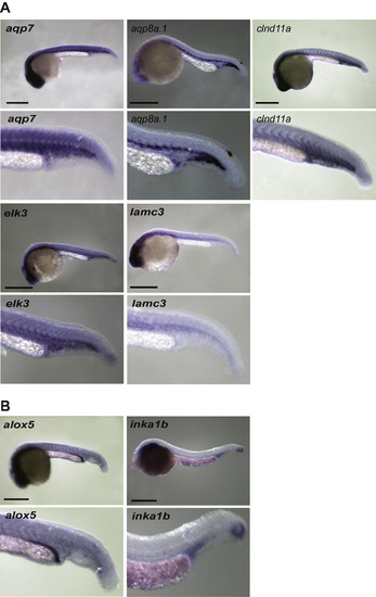
Remaining in situ hybridisation expression patterns in 24–28hpf embryos. (A) Vascular expression (B) Other. All embryos are lateral views with anterior to left. Scale bars indicate 500 μm. |
Expression Data
| Genes: | |
|---|---|
| Fish: | |
| Anatomical Terms: | |
| Stage: | Prim-5 |
Expression Detail
Antibody Labeling
Phenotype Data
Phenotype Detail
Acknowledgments
This image is the copyrighted work of the attributed author or publisher, and
ZFIN has permission only to display this image to its users.
Additional permissions should be obtained from the applicable author or publisher of the image.
Reprinted from Mechanisms of Development, 130(2-3), Cannon, J.E., Place, E.S., Eve, A.M., Bradshaw, C.R., Sesay, A., Morrell, N.W., and Smith, J.C., Global analysis of the haematopoietic and endothelial transcriptome during zebrafish development, 122-131, Copyright (2013) with permission from Elsevier. Full text @ Mech. Dev.