
Fig. 4
- ID
- ZDB-FIG-130228-1
- Publication
- Cerdà et al., 2002 - Molecular characterization of calymmin, a novel notochord sheath-associated extracellular matrix protein in the zebrafish embryo
- Other Figures
- All Figure Page
- Back to All Figure Page
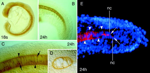
Localization of Calymmin in wild-type zebrafish embryos by whole-mount immunocytochemistry (A-D) and immunofluorescence microscopy (E). Views of embryos with rostral to the left (A-C), cross-section of labelled embryos (D), and cryostat section (E). A: 18-somite stage embryo showing staining within the developing notochord. B,C: Embryos at the pharyngula period (24 hours postfertilization [hpf]) showing Calymmin staining at the notochord sheath (C), which is less intense at the level of the tail (B). The arrowheads point to the notochord sheath, whereas the arrows indicate subcellular staining deposits close to the nucleus of the notochord cells. D: Cross-section of labelled embryos at the level of the trunk showing positive and specific immunolocalization of Calymmin in the notochord sheath. Note the high vacuolization of the notochord cells that are surrounded by the sheath. E: Immunofluorescence localization of Calymmin (red color) in the notochord cells located in the tail and anterior trunk of 24-hpf embryos. The nucleus of the notochord cells can be visualized by the Hoechst fluorescent dye (blue color). The arrows indicate the juxtanuclear localization of Calymmin within the notochord cells, likely in the Golgi apparatus, whereas the arrowheads point to the appearance of Calymmin at the notochordal sheath. nc, nucleus of the notochord cells. |
| Gene: | |
|---|---|
| Antibody: | |
| Fish: | |
| Anatomical Terms: | |
| Stage Range: | 14-19 somites to Prim-5 |